
CASE OF THE DAY 6 NOV
Question:
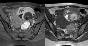
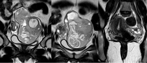
A 33-year-old women with pelvic pain.
What is the best diagnosis?
- Answer:
3) Twisted pedicle in the right side with right ovarian dermoid.
- Findings:
coronal T2-weighted MR images shows well-defined right ovarian mass with heterogeneous signal Intensity. Axial T2-fat sat and T1-non fat sat MR images demonstrate significant loss of signal intensity with-in the adnexal mass in the fat suppress image (ovarian dermoid). A whorled structure in right side represents a twisted vascular pedicle (ovarian torsion) .
- Discussion:
- Ovarian dermoid cystand mature cystic ovarian teratomaare terms often used interchangeably to refer to the most common ovarian neoplasm. These slow-growing tumors contain elements from multiple germ cell layers and are best assessed with ultrasound. The spectrum of sonographic features includes: diffusely or partially echogenic mass with posterior sound attenuation owing to sebaceous material and hair within the cyst cavity ,an echogenic interface at the edge of mass that obscures deep structures: the tip of the iceberg sign ,mural hyperechoic Rokitansky nodule (dermoid plug) , echogenic, shadowing calcific or dental (tooth) components ,the presence of fluid-fluid levels ,multiple thin, echogenic bands caused by the hair in the cyst cavity: the dot-dash pattern (dermoid mesh) ,color Doppler: no internal vascularity ,internal vascularity requires further workup to exclude a malignant lesion ,intracystic floating balls sign is uncommon but characteristic.
- Typically CT images demonstrate fat (areas with very low Hounsfield values), fat-fluid level, calcification (sometimes dentiform), Rokitansky protuberance, and tufts of hair. The presence of most of the above tissues is diagnostic of ovarian cystic teratomas in 98% of cases . Whenever the size exceeds 10 cm or soft tissue plugs and cauliflower appearance with irregular borders are seen, malignant transformation should be suspected.
- MR evaluation usually tends to be reserved for difficult cases but is exquisitely sensitive to fat components. Both fat suppression techniques and chemical shift artifact can be used to confirm the presence of fat.
- ovarian torsion occur in ~3-16% of ovarian teratomas and in general: considered the most common complication of dermoid.
- Rha, Sung E., et al. “CT and MR imaging features of adnexal torsion.” Radiographics 22.2 (2002): 283-294.
- Chang, Hannah C., Shweta Bhatt, and Vikram S. Dogra. “Pearls and pitfalls in diagnosis of ovarian torsion.” Radiographics 28.5 (2008): 1355-1368.
- Outwater, Eric K., Evan S. Siegelman, and Jennifer L. Hunt. “Ovarian teratomas: tumor types and imaging characteristics.” Radiographics 21.2 (2001): 475-490.
Courtesy of Behnaz Moradi,MD,Assistant Professor Department of Radiology, Yas HospitalTehran University of Medical Sciences.