
CASE OF THE DAY 4 NOV
Question:
A 50 years old woman with chronic disease and post biopsy hematoma.
Which kind of intervention has been done?
- Answer:
3) Transplant kidney AVF coiling.
- Findings:
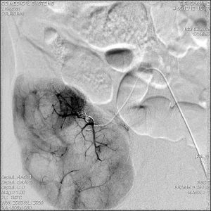
- DSA of Transplanted renal artery (early arterial phase) shows a micro catheter which is inserted into the feeding artery selectively. Selective DSA image of the feeding artery shows early filling of the renal vein suggesting an AV fistula, A small pseudoaneurysm is also visible.
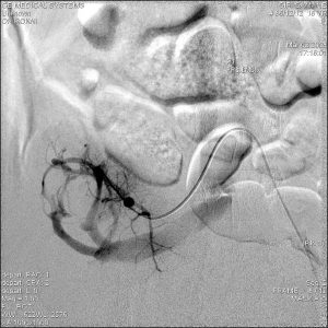
- DSA of Transplanted renal artery (late arterial phase) after trans arterial coil embolization shows complete occlusion of the AV fistula and pseudoaneurysm.
- Discussion:
- The most common cause of traumatic renal AV shunts is iatrogenic injury, especially percutaneous renal biopsy. Several studies reported incidences of 7.4%–11% after renal biopsy. A majority of traumatic renal AV shunts due to renal biopsy are asymptomatic and resolve spontaneously within 2 years, but some can be symptomatic and require interventional treatment. They are usually solitary, involving a single direct communication between the renal artery and adjacent vein, so-called traumatic AV fistulas. Pseudoaneurysms occasionally coexist with traumatic renal AV shunts.
- DSA has been the gold standard technique for the evaluation of renal AV shunts because of its high spatial and temporal resolution, which allows for the assessment of the detailed angioarchitecture and hemodynamics of renal AV shunts. However, recent advances in noninvasive imaging modalities, including color Doppler US, multi– detector row CT angiography, and magnetic resonance (MR) angiography, are also helpful in the diagnosis of renal AV shunts.
- Traumatic shunts are often associated with pseudoaneurysms and often located in the periphery of the kidney. Catheterization with a microcatheter is thus required for small feeders, and NBCA and/or coils are commonly used to achieve complete obliteration of both the renal AV shunt and the associated pseudoaneurysm.
- Maruno M, Kiyosue H, Tanoue S, Hongo N, Matsumoto S, Mori H, et al. Renal arteriovenous shunts: clinical features, imaging appearance, and transcatheter embolization based on angioarchitecture. Radiographics 2016;36(2):580–95.
- Raju DS, Rammurti S. Arteriovenous fistula following kidney biopsy. Indian J Nephrol. 2008;18:83–4.
Courtesy of Hadi Rokni,MD, Full Professor department of Radiology, Imam Khomeini Hospital Tehran University of Medical Sciences.