
CASE OF THE DAY 12 NOV
Question:
A 5 year old boy presents to the emergency room with nondescript abdominal pain.
What is your diagnosis?
|
|
|
Answer:
2) Pulmonary sequestration
Findings:
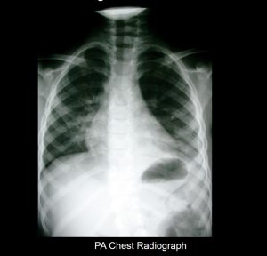
- The CXR demonstrates a retrocardiac paraspinal opacity and a small left pleural effusion.
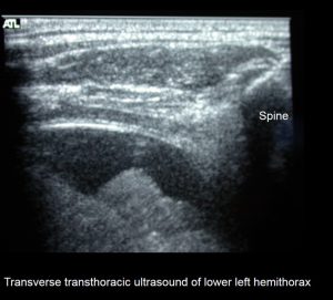
- Transthoracic ultrasound demonstrates a paraspinal mass and a small effusion.
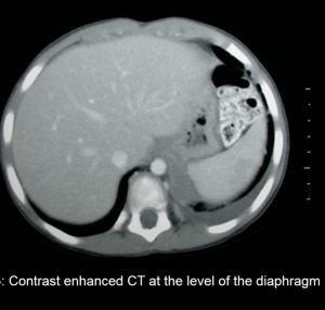
- The CT imaging also demonstrates a small mildly enhancing soft tissue mass and localizes it to the level of the left hemidiaphragm.
Discussion:
Pulmonary sequestration, also called accessory lung, refers to the aberrant formation of segmental lung tissue that has no connection with the bronchial tree or pulmonary arteries. It is a bronchopulmonary foregut malformation (BPFM).
There are two types:
- intralobar sequestrations
- venous drainage commonly occurs via the pulmonary veins but can occur through the azygous-hemiazygous system, portal vein, right atrium or inferior vena cava
- closely connected to the adjacent normal lung and do not have a separate pleura
- extralobar sequestrations
- venous drainage most commonly through the systemic veins into the right atrium (but is variable)
- separated from any surrounding lung by its own pleura
Overall, sequestration preferentially affects the lower lobes. 60% of intralobar sequestrations affect the left lower lobe, and 40% the right lower lobe. Extralobar sequestrations almost always affect the left lower lobe, however, ~10% of extralobar sequestrations can be subdiaphragmatic
Plain radiograph
- often show a triangular opacity in the affected segment
- may show cystic spaces if infected
- both ILS and ELS can rarely have air bronchograms as they may acquire a connection to the bronchial tree due to an infective process or, rarely, they can have foregut communication (esophagus or stomach) as part of hybrid lesion
Ultrasound
The sequestrated portion of the lung is usually more echogenic than the rest of the lung. On antenatal ultrasound, an extralobar sequestration may be seen as early as 16 weeks gestation and typically appears as a solid well-defined triangular echogenic mass .Color Doppler may identify a feeding vessel (in-utero cases) from the aorta. If the sequestration is subdiaphragmatic, it may appear as an echogenic intra-abdominal mass.
CT
- cross-sectional imaging frequently demonstrates the arterial supply by the descending aorta
- they may arise below the diaphragm in 20% of patients
- usually does not contain air unless infected
- 3D reconstructions can be particularly helpful in detecting
- anomalous arterial vessels
- concurrent anomalous veins
- differentiating between intralobar and extralobar sequestrations
Courtesy of Ali Hekmatnia,MD, Full Professor Department of Radiology Isfahan University of Medical Sciences.